| . |  |
. |
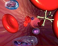
A research team from Oregon Health & Science University and the Portland Veterans Affairs Medical Center is demonstrating some of the world's first clinical applications for nanometer-size particles in the brain. The OHSU scientists have shown that an iron oxide nanoparticle as small as a virus can outline not only brain tumors under magnetic resonance imaging, but also other lesions in the brain that may otherwise have gone unnoticed, according to a study published in the journal Neuropathology and Applied Neurobiology. So named because of its billionth-of-a-meter proportions, the iron oxide nanoparticle, ferumoxtran-10, can be viewed as a contrast agent under MR for more than 24 hours, sometimes as long as five days, said the study's lead author, Edward Neuwelt, M.D., professor of neurology and neurological surgery, OHSU School of Medicine, and the Portland VA Medical Center. In a parallel study by Neuwelt and colleagues, to be presented in June to the American Society of Neuroradiology, ferumoxtran-10 also was found to provide a "stable imaging marker" during surgery to remove brain tumors, and it remains in the brain long enough for post-operative MR, even after surgical manipulation. The studies' findings have the potential to assist image-guided brain surgery and improve diagnosis of lesions caused by multiple sclerosis, stroke and other neurological disorders, in addition to residual tumors. "This is one of the first biologically specific nanoparticles to be used in clinical trials," said Neuwelt, director of OHSU's Blood-Brain Barrier Program, which studies ways of outwitting the brain's natural defense to treat people with brain tumors. "This is the first time it's been applied to look at inflammatory lesions in the brain, not just tumors. They're very interesting particles and they're safe." Study co-author Gary Nesbit, M.D., associate professor of diagnostic radiology and neurological surgery, and the Dotter Interventional Institute, OHSU, agreed the nanoparticles show potential for providing insight into a variety of brain pathologies. "We're learning that disease in the brain is a complex system that has active involvement with cells and membranes, so there's a lot of interest in other areas as well," Nesbit said. He said this early research on ferumoxtran-10 must be expanded to a larger group of patients to help scientists learn more about how the material reaches certain tumors and lesions, and why. "This contrast agent provides a completely different way of looking at enhancement patterns on MRI," Nesbit added. There are distinct differences in how tumors enhance with contrast agents. "The way the contrast agent gets there is completely different. What we need to find out is what the specific pattern of enhancement with this contrast agent means." Because ferumoxtran-10 can stay in brain lesions for days - it can be administered to patients 24 hours before surgery - and can image other, non-cancerous lesions, it has some advantages over gadolinium, a metal used as an MR contrast agent for 20 years and which must be administered just before surgery. But Neuwelt doesn't believe iron oxide nanoparticles will necessarily replace gadolinium as an imaging tool. "It will complement gadolinium, but not replace it," he said. "Gadolinium is the gold standard. But ferumoxtran-10 gives us additional information we can't get in some patients with gadolinium. Using both kinds of contrast agents, we can get better diagnostic information and that has the potential to improve the patient's outcome." In the Neuropathology and Applied Neurobiology study, which followed seven patients with primary and metastatic malignant tumors, researchers used Combidex, a ferumoxtran-10 manufactured by Advanced Magnetics Inc. of Cambridge, Mass. Each iron oxide nanoparticle is the size of a small virus and is much smaller than a bacterium but much larger than an atom or standard gadolinium contrast molecule, Neuwelt said.
"It's an iron oxide crystal surrounded with a carbohydrate or 'sugar' coating," Neuwelt said. This coating, called dextran, gives the particle a longer plasma half-life, allowing it to slowly slip through the blood-brain barrier, or "Anytime there's an injury, it induces inflammation," Neuwelt said. Ferumoxtran-10 is "taken up by inflammatory cells in the brain. You can see them in stroke and MS, you can see them in tumors. Gadolinium is basically the size of a large atom and does not enter cells, while this contrast agent is the size of a small virus and does enter cells." In addition, ferumoxtran-10 can be detected with an iron stain in the tissue removed by biopsy or surgery, allowing physicians to see it in brain tissue samples under a microscope. "Unlike any other MR contrast agent, you can compare the images from an MR scan with the tissue taken out at surgery," Neuwelt said. And it's relatively safe when diluted and administered as an infusion, although it can cause an allergic-type reaction when administered too quickly, he said. Neuwelt says he first learned about iron oxide nanoparticles 11 years ago at a National Institutes of Health conference on new medical imaging techniques. "I heard this talk, and they were using it for liver and lymph nodes and I said to myself, 'Well, we ought to be able use it for the brain as well,'" he recalled. Soon after, in April 1994, Neuwelt and colleagues published a study in the journal Neurosurgery showing that iron oxide nanoparticles could be delivered across the blood-brain barrier to the brain cells of rats and could be seen with MR. "Once we realized that iron oxide nanoparticles had unique pharmacology and a very long half-life in blood, we thought this might be a good imaging modality, not just to see where viruses went, but where inflammatory regions in the brain are," Neuwelt said. Robert Quencer, M.D., chairman of the Department of Radiology at the University of Miami School of Medicine, and chief of radiological services at Jackson Memorial Hospital in Miami, is editor-in-chief of the American Journal of Neuroradiology, which published one of the first articles on the use of nanoparticles in imaging human brain tissue in 2002. Quencer called Neuwelt's study "significant" because ferumoxtran-10 stays around longer than gadolinium, allowing doctors to view the affected tissue throughout the surgical process by intraoperative MR. And exploration of the mechanism by which it's accumulated in cells may provide more insight about tumors and other areas where reactive cells are found. "There are a lot of possibilities," he said. "One area would be in spinal cord tumors. There's no reason to suspect the uptake would be different in the spine. And it could be very valuable in guiding a biopsy." Neuwelt's collaborators included Nesbit; Peter Varallyay, M.D., instructor in diagnostic radiology; Attila Bago, M.D., former research instructor in neurology; Leslie Muldoon, Ph.D., assistant professor of cell and developmental biology, and neurology; and Randal Nixon, M.D., assistant professor of pathology. Related Links Oregon Health & Science University SpaceDaily Search SpaceDaily Enigmatic X-Ray Sources May Point To New Class Of Black Holes  Boston - Mar 08, 2004
Mysterious, powerful X-ray sources found in nearby galaxies may represent a new class of objects, according to data from NASA's Chandra X-ray Observatory. These sources, which are not as hot as typical neutron-star or black-hole X-ray sources, could be a large new population of black holes with masses several hundred times that of the sun.
|
| ||||||||||
| The content herein, unless otherwise known to be public domain, are Copyright 1995-2016 - Space Media Network. All websites are published in Australia and are solely subject to Australian law and governed by Fair Use principals for news reporting and research purposes. AFP, UPI and IANS news wire stories are copyright Agence France-Presse, United Press International and Indo-Asia News Service. ESA news reports are copyright European Space Agency. All NASA sourced material is public domain. Additional copyrights may apply in whole or part to other bona fide parties. Advertising does not imply endorsement, agreement or approval of any opinions, statements or information provided by Space Media Network on any Web page published or hosted by Space Media Network. Privacy Statement All images and articles appearing on Space Media Network have been edited or digitally altered in some way. Any requests to remove copyright material will be acted upon in a timely and appropriate manner. Any attempt to extort money from Space Media Network will be ignored and reported to Australian Law Enforcement Agencies as a potential case of financial fraud involving the use of a telephonic carriage device or postal service. |